PERITONEAL FOLDS

Double layered peritoneal folds, variously named as ligaments, omenta and mesenteries, connect the intraperitoneal organs to the abdominal wall. Some of these ligaments contain blood vessels and lymph nodes. while others are avascular.
EMBRYOLOGY DEVELOPMENT OF PERITONEAL FOLDS:

The fold can be best understood by recapitulating the embryology of the gut; the developing gut is divisible into 3 parts forgut, midgut and hindgut; each part has its own artery which is a ventral branch of the abdominal aorta. The coeliac artery supplies the forgout, the superior mesenteric artery supplies the midgut, and the inferior mesenteric artery supplies the hindgut.
Apart from some other structures, the foregut forms the oesophagus, the stomach, and the upper part of the duodenum up to the opening of the bile duct. the midgut forms the rest of the duodenum, the jejunum, the ileum, the appendix, the caecum, the ascending colon and the right two-third of the transverse colon, the Hindgut forms the left one- third of the transverse colon, the descending colon, the sigmoid colon,proximal part of the rectum. the anorectal canal forms dister part of the rectum and the upper part of the anal Canal up to the pectinate line.
The abdominal part of the foregut is suspended by mesenteries both ventrally and dorsally. The ventral mesentery of the forgot is called the ventral mesogastrium, and the dorsal mesentery is called dorsal mesogastrium (gastrium mean stomach)
The ventral mesogastrium becomes divided by the developing liver into a ventral part and a dorsal part.
the ventral part forms the ligaments of the liver, namely:
The falciform ligament
The right and left triangular ligaments, and
The superior and inferior layers of the coronary ligament.
The dorsal part of the ventral mesogastrium forms the lesser omentum.
The fate of the dorsal Mesogastrium is as follows.
The greater and caudal part of the dorsal mesogastrium becomes greatly elongated and forms the greater omentum.
The spleen develops in relation to the cranial part of the dorsal and ventral parts. The ventral part forms the gastrosplenic ligament while the dorsal part forms the gastrosplenic ligament while the dorsal part forms the lienorenal ligament.
The cranial most part of the dorsal mesogastrium forms the gastrophrenic ligament.
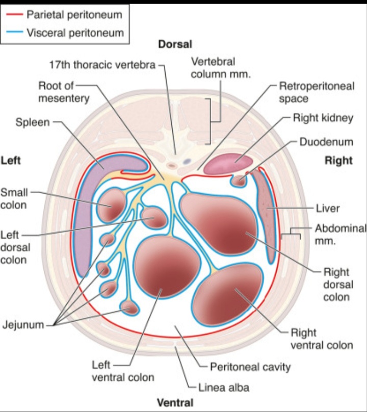
The midgut and hindgut have only a dorsal mesentery, which forms the mesentery of jejunum and ileum, the mesoappendix, the transverse mesocolon and the sigmoid mesocolon. The mesentery of the duodenum, the ascending colon, the descending colon and the rectum are lost during development.
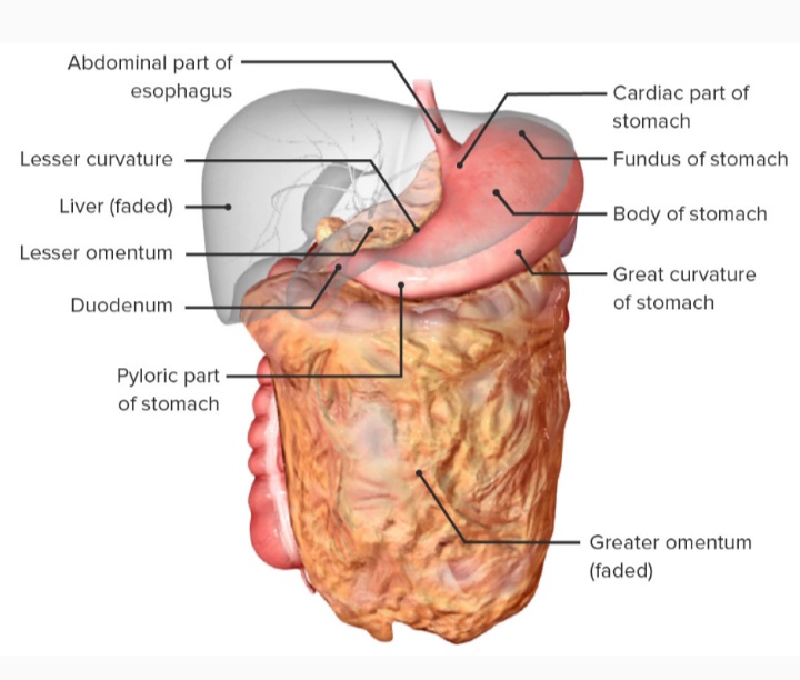
GREATER OMENTUM
The greater Momentum is a large fold of peritoneum which hangs down from the greater curvature of the stomach like an apron and covers the loop of intestine to a varying extent. It is made up of 4 layers of peritoneum all of which are Fused together to form a thin fenestrated membrane containing variable quantities of fat and small arteries and veins.
ATTACHMENTS
The anterior two layers descend from the greater curvature of the stomach to a variable extent and fold upon themselves to form the posterior two layers which ascend to the anterior surface of the head, and the anterior border of the body of the pancreas. The folding of the omentum is such that the first layer becomes the fourth layer and the second layer becomes the third layer. In its upper part of the fourth layer is partially fused to the anterior surface of the transverse colon and of the transverse mesocolon. The part of the Peritoneal cavity called the lesser sac between the second and third layers gets obliterated, except for about 2.5cm below the greater curvature of the stomach.
Contents
The right and left gastroepiploic arteries (also known as gastroomental) provide the sole blood supply to the greater omentum. Both are branches of the celiac trunk. The right gastroepiploic artery is a branch of the gastroduodenal artery, which is a branch of the common hepatic artery, which is a branch of the celiac trunk. The left gastroepiploic artery is the largest branch of the splenic artery, which is a branch of the celiac trunk. The right and left gastroepiploic arteries anastomose within the two layers of the anterior greater omentum along the greater curvature of the stomach.
It is often laden with fat.
Functions
It's a storehouse of fat.
It protects the peritoneal cavity against infection because of the presence of macrophages from small, dense patches, known as milky spots, which are visible to the naked eye.
It also limits the spread of infection by moving to the site of infection and sealing it off from the surrounding areas. On this account, the greater momentum is also known as the policemen of the abdomen.
CLINICAL SIGNIFICANCE
Surgical removal:-
Omentectomy refers to the surgical removal of the omentum, a relatively simple procedure with no documented major side effects, that is performed in cases where there is concern that there may be spread of cancerous tissue into the omentum. Examples for this are ovarian cancer and advanced or aggressive endometrial cancer as well as intestinal cancer and also appendix cancer. The procedure is generally done as an add-on when the primary lesion is removed.
Omental flap:-
The greater omentum may be surgically harvested for reconstruction of the thoracic wall. It has also been used experimentally to reinforce bioengineered tissues transplanted to the surface of the heart for cardiac regeneration.
The greater omentum forms a partition between the supracolic and 6infracolic compartments of the greater sac.
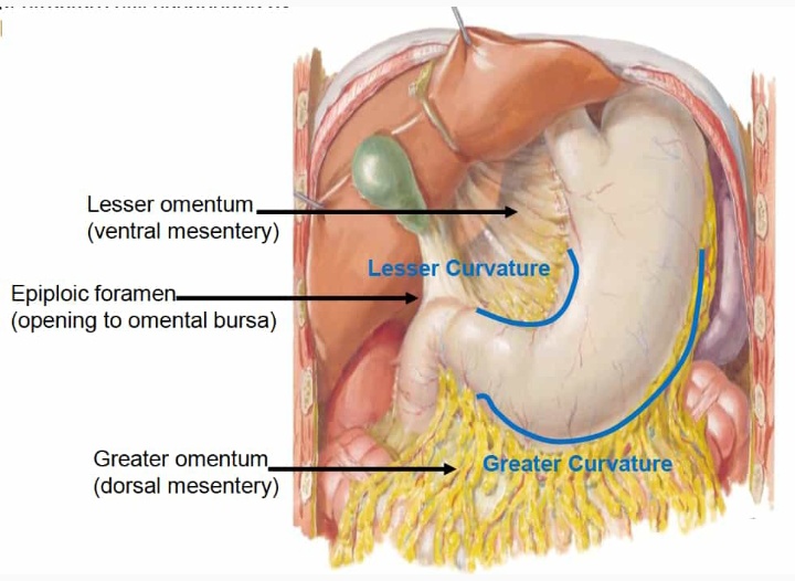
LESSER OMENTUM
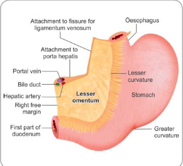
This is a fold of peritoneum which extends from the lesser curvature of the stomach and the first 2cm of the duodenum to the liver. The portion of the lesser omentum between the stomach and the liver is called the hepatogastric ligament and the portion between the duodenum and the liver is called the hepatoduodenal ligament. Behind the lesser omentum, there lies a part of the lesser sac. The lesser omentum has a free right margin behind which there is the epiploic foramen .The greater and lesser sac communicate through this foramen.
Attachments
Inferiorly, the lesser omentum is attached to the lesser curvature of the stomach and to the upper border of the first 2cm of the duodenum. Superiorly, it is attached to the liver, the line of attachment being in the form of an inverted 'L'. The vertical limb of the 'L' is attached to the bottom of the fissure for the ligamentum venosum, and the horizontal limb to the margins of the porta hepatis.

Contents
The right free margin of the lesser omentum contains:
The proper hepatic artery:,
The portal vein:,
The bile duct:,
Lymph nodes and lymphatics:, and
The hepatic plexus of nerves, all enclosed in a perivascular fibrous sheath.
Along the lesser curvature of the stomach and along the upper border of the adjoining part of the duodenum,it contains:
The right gastric vessels
The left gastric vessels
The gastric group of lymph node and lymphatics
Branches from the gastric nerve.
CLINICAL ANATOMY
ASCITES- collection of free fluid in the peritoneal cavity is known as Ascites. Common causes of ascites are cirrhosis of the liver, tubercular peritonitis,congestive heart failure, and malignant infiltration of the peritoneum. Veins also get prominent in cirrhosis of the liver.
PARACENTESIS- fluid from the peritoneal cavity may be removed by puncturing the abdominal wall either in the median plane midway between the umbilicus and pubic symphysis, or at a point just above the anterior superior iliac spine. The procedure is called paracentesis. The urinary bladder must be emptied before the procedure.
PERITONITIS- inflammation of the peritoneum is called peritonitis.it may be localized when a subjacent organ is infected:, or maybe generalized. The latter is a very serious condition.
PNEUMOPERITONEUM- The presence of air in the peritoneal cavity is called pneumoperiyonium.it may occur after perforation of the stomach and intestines.
LAPAROSCOPY- Laparoscopy is examination of the peritoneal cavity under direct vision using an instrument called laparoscopy.
Opening up the abdominal cavity by a surgeon is called laparotomy.
FUNCTION OF PERITONEUM
Movement of viscera: the chief function of the peritoneum is to provide a slippery surface for free moments of abdominal viscera. This permits peristaltic movement of the stomach and intestine, abdominal movement during respiration and periodic change in the capacity of hollow Viscera associated with their filling and evacuation. The efficiency of the intestine is greatly increased as a result of the wide range of mobility that is possible because the intestines are suspended by a large fold of peritoneum.
Protection of viscera: The peritoneum contains various phagocytic cells which against infection Lymphocytes present in normal peritoneal fluid provide both cellular and humoral immunological defense mechanisms. The greater momentum has the power to move towards the site of infection and to Seal them thus preventing spread of infection. For this reason, the Greater omentum is often designated as the ' policeman of the abdomen'.
Absorption and dialysis: the mesothelium acts as a semipermeable membrane across which Fluids and small molecules of various solutes can pass. Thus, the peritoneum can absorb fluid effusions from the peritoneal cavity. water and crystalloids are absorbed directly into the blood capillaries, whereas colloids pass into lymphatics with the aid of phagocytes. The greater Absorptive power of the upper abdomen or sUbphrenic area is due to its large surface area and because respiratory movements aid absorption.
Therapeutically considerable volume of fluid can be administered through the peritoneal route. Conversely, metabolites, like urea can be removed from the blood by artificially circulating fluid through the peritoneal cavity. This procedure is called peritoneal dialysis.
Healing power and adhesions: The mesothelium cell of the peritoneum can transform into fibroblast which promotes healing of wounds.
Storage of fat: peritoneal fold are capable of storing large amount of fat, particularly in obese person.
Provides passage for nerves, vessels and lymphatics to and from the suspended viscera.
THANK YOU


Wow!!
ReplyDeleteThat's great ✌✌😍😍