BLOOD
Blood is a special connective tissue consisting of a fluid matrix, plasma and formed elements.
PHYSICAL APPEARANCE
https://kareenalohia232406.blogspot.com/2022/07/memory-what-is-infornation-processing.htmlBlood is an opaque, mobile fluid connective tissue, mesodermal in origin. It is somehow sticky, and slightly heavier than water bulk for bulk (specific gravity 1.06).It has saltish taste and a mild alkaline reaction PH is 7.4.Its osmotic pressure at 37 degrees celsius is about 7.6 atmosphere. It is bright red when oxygenated and purple when deoxygenated.
COMPOSITION
BLOOD consists of a watery fluid called plasma containing certain floating bodies termed formed elements. The plasma and formed volume of the blood respectively.
PLASMA:- The plasma is a straw coloured, viscous fluid constituting nearly 55 percent of the blood. It is a complex mixture which is in dynamic equilibrium with the intracellular fluid bathing the cells and the intracellular fluid present within the cells.
It constantly takes up and loose materials as it flows through the capillaries, yet it has a constant chemical composition. It consists of about 90% water 1% inorganic salts in true solution and 7 or 8% proteins in colloidal state. The plasma is formed by food materials, waste products, dissolved gases, regulatory substances, anticoagulant cholesterol and antibodies. These substances do not form an integral part of the plasma as they enter and leave at intervals. They are being carried by the plasma from one place to another in the body.
Proteins:- The plasma contains a number of proteins : Serum albumin, globulins, prothrombin and fibrinogen. The plasma proteins serve many functions :
They serve as acid -base buffers, they maintain PH of the blood by neutralizing acid and base.
Albumins and globulins maintain osmotic pressure of the plasma so that the letter may retain water. Fall in the level of plasma proteins causes excessive filtering of water from the blood into the tissues. This may produce oedema (swelling) of hands and feet in persons taking protein -deficient diet.
Plasma proteins transport certain material in combination with them. Thyroxine is bound to albumin or a specific globulin, insulin is combined with globulin, fatty acids are joined to albumen for transport in the plasma.
Some globulin called immunoglobulins (Ig), form protective proteins, termed antibodies, in response to the entry of foreign agents, the antigens,into the body. The antibodies inactivate the antigens
Prothrombin and fibrinogen play a role in blood clotting.
INORGANIC SALTS
The inorganic salts occur in the plasma as ions. Sodium and chloride are the principal cation and anion of the plasma. The anions bicarbonate and phosphate, and the cation potassium,magnesium,calcium,
Iron and manganese occur in smaller amounts. The inorganic salt present in the plasma as dissolved ions are often called blood (plasma) electrolytes.
https://kareenalohia232406.blogspot.com/2022/03/epithelial-tissue.html
FOOD MATERIALS
The food materials present in the plasma are glucose, amino acid, fatty acids and triglycerides. Their amount depends upon the digestion of food in the alimentary canal. Normally an adult person has 80 to 100mg.of glucose per 100 ml of blood 12 hours after a meal.
If blood sugar exceeds 180mg., glucose is excreted in the urine, causing the disease diabetes mellitus or hyperglycemia.
Fall in blood sugar is called hypoglycemia.
waste products :- The waste products found in the plasma are urea, uric acid, ammonia and creatinine. These are removed by the kidneys. Their excess causes a toxic effect called uraemia.
Dissolved Gases :- small amounts of oxygen, carbon dioxide and nitrogen are dissolved in the plasma.
Regulatory Substances :- These include hormones, vitamins and enzymes.
Anticoagulant :- A natural strong anticoagulant present in the plasma is a heteropolysaccharide named antiprothrombin, or heparin. It checks clotting of blood in uninjured blood vessels by preventing the conversion of prothrombin into thrombin. It is produced in the liver.
Cholesterol :- Liver synthesizes cholesterol and releases it into the blood. It is also absorbed into the blood from the food, such as egg, digested in the intestine. It provides material to the tissue cells for the synthesis of membrane lipids, vitamin D, steroid hormones and bile salt. Cholesterol normally ranges from 50 to 180mg. Per 100 ml. Of blood. Rise in the level of cholesterol in the blood may cause heart trouble.
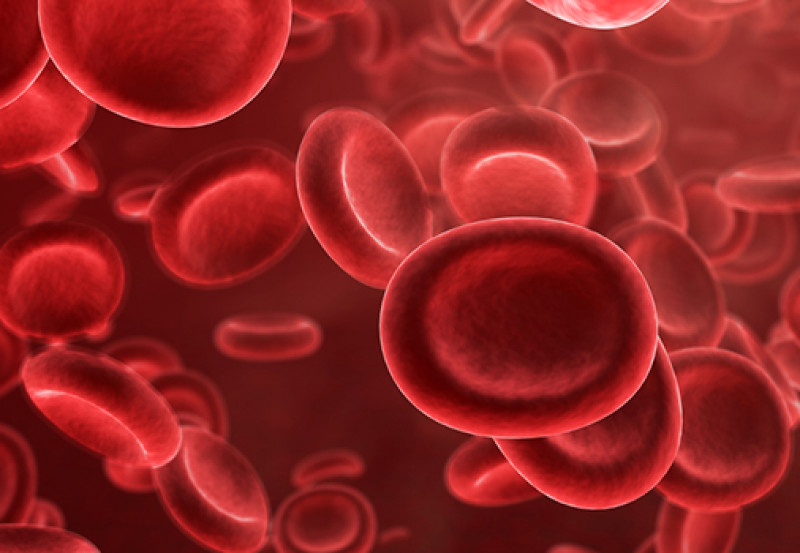
RED BLOOD CORPUSCLES (RBC)
The red blood corpuscles are the most numerous formed elements of the blood. They are the most abundant cells in the human body. The unique feature of the RBCs is the presence of a red, oxygen - carrying pigment, the haemoglobin, in their cytoplasm.
Shape :- The shape of RBCs varies in different vertebrate classes. In fishes, amphibians, reptiles and birds, they are oval, biconvex and nucleated. In mammals, they are circular, biconcave, denuclated discs. Their central part is thinner than the Margin. This shape provides. flexibility and results in a 20to 30% increase in surface area as compared to a sphere. This favours quick diffusion of gases.

Size :- Human RBCs are smaller than the white corpuscles.
Number :- The RBCs are far more numerous than the WBCs. A normal healthy adult man and woman have about 5.5 and 4.5 million RBCs per cubic millimeter of blood respectively. This is called the total RBCs count. Anemia may be caused by loss of blood (haemorrhage), destruction of RBCs (haemolysis).
The RBCs count increase during exercise to meet the increased demand of oxygen and at high altitudes to cope with the low oxygen content of the air.
An abnormal rise in RBCs count is called polycythemia.
Decrease in the number of red blood corpuscles, termed erythrocytopenia, cause oxygen shortage in the blood and tissue. The oxygen shortage stimulates the kidneys cells to secrete a hormone,called erythropoietin,into the blood.
This hormone, in turn, stimulates the bone marrow to increase the production of red blood corpuscles. Addition of red corpuscles increases the oxygen -carrying capacity of the blood. As the blood's oxygen level becomes normal, secretion of erythropoietin stops and the production of red blood corpuscles return to normal.
Colour :- The RBCs look yellowish when seen singly and red viewed in bulk. They impared red colour to the blood. The colour is due to the solution of iron containing pigment, haemoglobin in them.
HAEMOGLOBIN

Hemoglobin is a conjugated protein. It consists of a basic protein globin joined to a non protein group heme, hence the name haemoglobin. Heme is an iron - porphyrin ring.
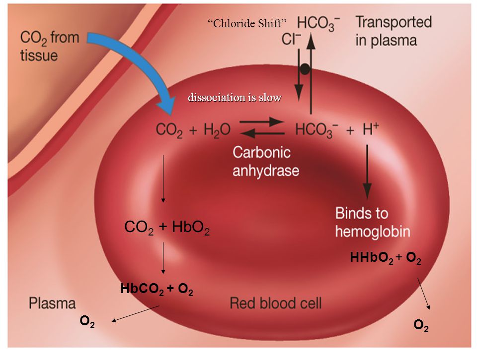
A hemoglobin molecule is a complex of 4 heme molecules joined with 4 globin molecules. There is about 15mg. Of haemoglobin in 100ml. of blood. In the lungs,due to high partial pressure of oxygen, haemoglobin takes up oxygen and changes to bright red oxyhaemoglobin. The letter carries 4 oxygen molecules loosely joined to 4Fe ++ ions. In the tissue, due to low partial pressure of oxygen, haemoglobin splits into oxygen and deoxyhaemoglobin. In this way, the RBCs carry oxygen from the lungs to the tissues.
RBCs also carry carbon dioxide from the tissue to the lungs for elimination. It is transported in two forms: mainly in combination with the water of RBC, forming bicarbonate ions.
Structure :- A red blood corpuscle is bound by an elastic and semi permeable plasma membrane. This enables it to squeeze through capillaries having a diameter less than their own.It loose plasticity in sickle cell anemia. In this disorder, the RBCs block the capillaries leading to grave consequences. An erythrocyte contains homogeneous cytoplasm which lose the nucleus, endoplasmic reticulum, mitochondria, ribosome and centrioles during the development of the corpuscle.This gives double advantage. The corpuscles have more space to hold haemoglobin. It's oxygen consumption is very low due to lack of organelles so that it can supply more oxygen carried by haemoglobin to the tissue cells. Red blood corpuscles cannot reproduce or carry out cellular metabolism due to lack of organelles. Besides haemoglobin, a red corpuscles also contains several inorganic ions, including those of sodium, potassium, calcium, magnesium, chloride and phosphate. The adult red blood corpuscles of mammals are described as nucleated (denuclated) as when young, they have a nucleus that later disappeared.
Formation :- Formation of red corpuscles Is called erythropoiesis. It occurs in the liver and spleen in the fetus and in red bone marrow after birth. Protein and iron are components of haemoglobin, and vitamin B12 and folic acid stimulate erythropoiesis. Excess RBCs are stored in the spleen.
Life span and disposal :- Human RBCs remain functional in the blood for about 120 days. The worn out RBCs are destroyed by phagocytosis in the blood itself and in the spleen and liver in particular. Their iron is returned to the red bone marrow for reuse in the synthesis of fresh haemoglobin. Their pigment is degraded to yellowish pigment bilirubin which is excreted in bile. The pale yellow color of the plasma is mainly due to bilirubin. If bilirubin Is not excreted fully, the skin and mucous membranes of the person become yellowish. This disorder is called jaundice.
Special property :- In resting (drawn) and slow flowing blood, the RBCs form piles called rouleaux by adhering together due to surface tension.
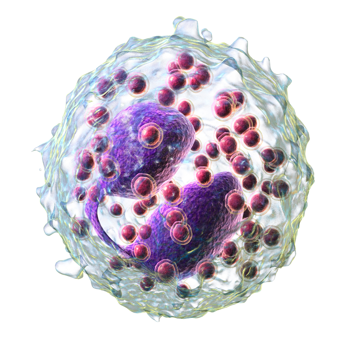
WHITE BLOOD CORPUSCLES
The white blood corpuscles (formed elements) lack haemoglobin.

Shape :- The WBCs are round or irregular cells.They can change their Shape and are capable of amoeboid movement. This enables them to squeeze out of capillaries into the tissues. This process is called diapedesis.
Size :- The WBCs are mostly larger than the red corpuscles. They range in size from 12 to 20 micrometers.
Number :-The WBCs are far fewer than the RBCs. Their number varies from 5,000 to 10,000per cubic millimeter of blood. This number is the total count of WBCs. It may increase or decrease abnormally in certain conditions. Rice in WBCs count is called leukocytosis. It is a physiological response to fight infections(e.g pneumonia), fall in WBCs count is termed leukopenia. It occurs in conditions such as Folic Acid deficiency, infection of AIDS virus. WBCs count is useful in diagnosing disease.
Colors :- The WBC are colourless.
Structure :- The leukocytes are nucleated cells. Their cytoplasm contain mitochondria, hotline apparatus and centrioles besides other organelles.
Formation :- Formation of leukocytes is called leukopoiesis. It occurs in lymph nodes, spleen, thymus and red bone marrow.
Life span and disposal :- The leukocytes survive for a few (3-4) days only in the blood.
The WBCs are two types:-
Granular leukocytes
Agranular leukocytes
Agranulocytes These leukocytes lack granules in the cytoplasm and have non lobed, rounded or oval nucleus. Agranulocytes are called mononuclear cells. They have two subtype :-
Monocytes
Lymphocytes
The monocytes arise in the bone marrow. The B and T lymphocytes are produced in the bone marrow and thymus respectively. And mature in spleen and lymph nodes. Formation of granulocytes is termed granulopoiesis.
Monocytes :- these are the largest of all types of leukocytes; they have a large Subrounded or been shaped nucleus and a good amount of cytoplasm. They are very motile, are phagocytic and scavenger in action and engulf bacteria and cellular debris at injured sites. Generally they charge into macrophages after entering tissue spaces.

Lymphocytes :-These are about the size of the red corpuscles. They have a very large, rounded nucleus and scanty cytoplasm. They are non motile and non phagocytic. They secrete antibodies to destroy microbes and their toxin, reject grafts and kill tumour cells. They also help in healing of injuries. The lymphocytes may differentiate into 2 main types:

B lymphocytes
T lymphocytes
Granulocytes :- These leukocytes contain granules in the cytoplasm and have lobed nucleus. They are produced in the red bone marrow. Their formation is called granulopoiesis. They have 3 subtype :
Basophils
Eosinophiles
Neutrophils
Basophiles :- These take up basic stains such as methylene blue. They are fairly large and have nearly S-shaped nuclei and a few coarse granules. Granules contain histamine. The basophils release histamine and heparin by exocytosis into the blood.

Eosinophils :- These stain with acidic dyes such as eosin. They are also fairly large and have bilobed nucleus and abundant coarse granules. The eosinophiles have antihistamine properties. Their number increases in people with allergic conditions such as asthma or hay fever. They also help in dissolving blood clots.
Neutrophiles :-These stain equally well with both acidic and basic dyes. They are quite large and have many nucleus and abundant fine, azurophilic granules. The eosinophils are phagocytic in action. They engulf microbes. They are chemotactically attracted to bacteria peptidases.
.png)
PLATELETS

The platelets (formed elements) also lack haemoglobin.
Shape :- The platelets are rounded or oval, disk like bodies but quickly become stellate in extracted blood.
Size -: The platelets are the smallest formed element of the blood.
Number :- The platelets are fewer than the red corpuscles and more than the white blood corpuscles in Number. There are about 250,000 platelets in a cubic millimeter of blood. Increase and decrease in the number of platelets is known as thrombocytosis and thrombocytopenia respectively.
Formation :- The platelets are formed in the red bone marrow. The formation is known as thrombopoiesis.
Structure :- The platelets are flat, non nucleated fragments of giant cells called megakaryocytes of bone marrow, rather than true cells. They are bound by a membrane and contain a few organelles and secretory granules in the cytoplasm. They have at the center a group of basophilic granules, which give the appearance of a nucleus. At the site of injury, the platelets release platelet factor or thromboplastin that help in blood clotting.

Life span and disposal :- The platelets survive for 3-7 days only. They are disposed of by phagocytosis in the blood itself.
No comments:
Post a Comment