Sunday, February 27, 2022

BRYOPHYTES AND PTERDOPHYTES FULL INFORMATIVE NOTES

 BRYOPHYTES 

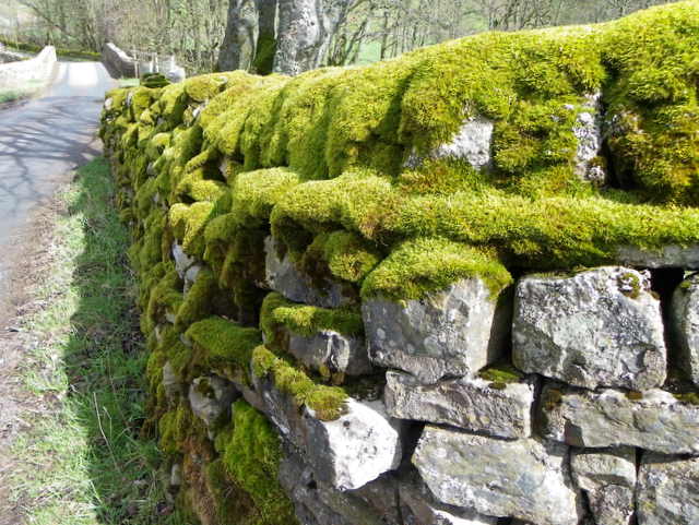
Bryophytes include the various mosses and liverwort that are found commonly growing in moist shaded areas in the hills.


  • Bryophytes are also called amphibians of the plant kingdom because these plants can live in soil but are dependent on water for sexual reproduction.

  • They usually occur in damp, humid and shaded localities.

  • They play an important role in plant succession on bare rock /soil. 

  • The plant body of bryophytes is more differentiated than that of algae. It is thallus-like and prostrate or erect, and attached to the substratum by unicellular or multicellular rhizoid.

  • They lack true roots, stems or leaves. 


  • They may possess root-like, leaf-like or stem-like structures.

  • The main plant body of the bryophyte is haploid. It produces gametes, hence is called a gametophyte.


  • The sex organs in bryophytes are multicellular. The male sex organ is called antheridium. They produce biflagellate antherozoids.The female sex organ is called archegonium is flask -shaped and produces a single egg. 

  • Theantherozoid are released into water where water where they come in contact with archegonium. 

  • An authrozoids fuses with the egg to produce the zygote. Zygotes do not undergo reduction division immediately. They produce a multicellular body called a sporophyte. 

  • The sporophyte is not free living but attached to the photosynthetic gametophyte and drives nourishment from it. 

  • Some cells of the sporophytes undergo reduction division (meiosis) to produce haploid spores. These spores germinate to produce gametophyte

Economic importance of Bryophytes 


  1. Soil conservation :- Mosses grow in dense mates over the soil surface. They bind the soil particles and prevent soil erosion by running water. 

  2. Formation of soil :- Mosses along with lichen play a very important role in the formation of soil over the bare rocky surface.  They grow on rock and add organic matter to the substratum after their death . It makes the rock surface suitable for the growth of higher plants. 

  3. Peat :-species of sphagnum, a moss, provide peat that have long been used as fuel,  and as packing material for trans - shipment of living material because of their capacity to hold water. 


LIVERWORTS


  • Liverworts are small, green, terrestrial plants. 


  • They do not have true roots, stems or leaves. 

  • They have an above ground leaf like structure, know as thallus, and an underground structure,known as a rhizoid 

  • Liverwort grow usually in moist, shady habitats such as banks of streams, marshy ground, damp soil, bark of trees and deep in the woods.

  • The plant body of liverwort is thalloid as in marchantia. The leafy members like porella have tiny leaf-like appendages in two rows on the stem-like structure.

  • Asexual reproduction occurs by means of fragmentation or by the formation of specialized structures called gemmae.


  • Gemmae formation is an important form of sexual reproduction in many species of liverwort and mosses.


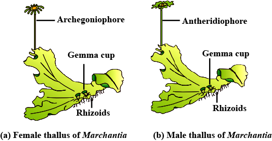
  • During sexual reproduction the sex organs antheridia (male) and archegonuia (female) are produced either on the same (Riccia) Or on different thallus (marchantia). 

  • In marchantia sex organ are Present on stalked recetcles. The male sex organ are present on antheridiophore and female sex organ are borne on the archegoniophore. 

  • Fusion of gamete result in the formation of zygote which develops into an embryo. 

  • The embryo in turn develops into sporophytes (diploid) .

  • The sporophytes are differentiated into foot, seta and capsule. Within the capsule, the spore mother cells undergo meiosis to produce the haploid spores. Spores on liberation germinate into the haploid free -living gametophyte. 


Mosses 


  • The predominant stage of the life cycle of a moss is the gametophyte which consists of protonema and leafy stage.

  • The plant body is a leafy gametophyte which has multicellular and branched rhizoids.


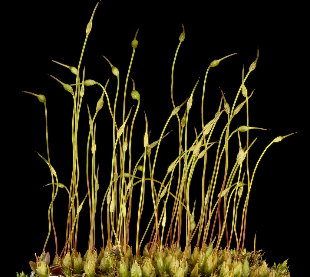
  • They consist of upright, slender axis bearing spirally arranged leaves.This stage bears the sex organs.

  • In sexual reproduction, the sex organs are present on the same plant but on different branches. 

  • Each antheridium produces a number of flagellate antherozoids. 

  • Each archegonium produces a fertile egg. Fusion Of gamete with the help of water leads to the formation of zygote. 

  • The zygote develops into a sporophyte which is differentiated into foot, seta and capsule. 


  • The capsule encloses two spore sacs, where spores are formed by meiosis. 

  • The mosses have an elaborate mechanism of spore dispersal.

  • Thespore on liberation germinates into a creeping,green, branched and frequently filamentous in mosses is by fragmentation and budding in the secondary protonema. 

  • The leafy stage develops from the secondary protonema as a lateral bud. 

Examples : Funaria, polytrichum and sphagnum .


PTE PTERIDOPHYTA

  • The pteridophyta include horsetail and ferns. Pteridophyta are used for medicinal purposes and as soil -binders. 

  • They are also frequently grown as ornamentals.

  • Evolutionary, they are the first terrestrial plants to possess vascular tissue - Xylem and phloem. 

  • The pteridophytes are found in cool, damp and shady places though some may flourish well in Sandy - soil conditions. 

  • In pteridophytes, the main plant body is a sporophyte which is differentiated into true root,stem and leaves. 

  • These organs possess well - differentiated vascular tissue. 

  • The live In pteridophytes are small (microphylls) as in selaginella or large (Macrophylls) as in ferns.

  • The sporophytes bear sporangia that are suspended by leaf-like appendages called sporophylls. 

  • In Some cases sporophyll may form distinct compact structures called strobili or cones (selaginella, equisetum) 

  • The sporangia produce spores by meiosis in spore mother cells. 

  • The spore germinates to give rise to inconspicuous, small but multicellular free living, mostly photosynthetic thalloid gametophytes called prothallus. 

  • These gametophytes require cool, damp, shady places to grow. Because of this specific restriction to narrow geographical requirement and need for water for fertilisation, the spread of living pteridophyta is limited and restricted to narrow geographical regions. 

  • The gametophytes bear male and female sex organs called antheridia and archegonia,respectively.

  • Water is required for transfer of antherozoids - the male gamete released from the antheridia,to the mouth of archegonium. 

  • Fusion of male gamete with the egg present in the archegonium results in the formation of zygote. 

  • Zygote thereafter produces a multicellular well- differentiated saprophyte which is the dominant phase of the pteridophytes. 

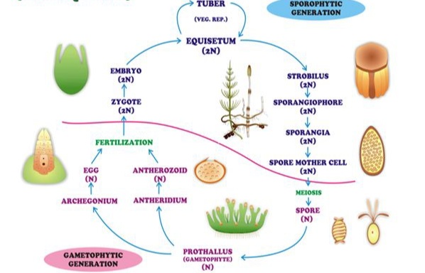
Life cycle of homosporous pteridophytes Examples : eqistem ,psilotum and lycopodium. 

  • In majority of the pteridophytes all the spores are of similar kinds :such plants are closed homosporous.


  •  selaginella and salvinia which produce two kinds of spores, macro (large) and micro (small) spores, are known as heterosporous

  • The megaspore and microspore germinate and give rise to female And male gametophytes, respectively. 

  • The female gametophytes in these plants are retained on the parent sporophytes for variable periods. 

  • The development of the zygote into young embryos takes place within the female gametophytes. 

Life cycle of heterosporous pteridophytes Example : Selaginella, marselia and salvinia. 

  • This event is a precursor to the seed habit considered an important step in evolution. 


The pteridophyta are further classified into four classes :


  • Psilopsida- Example : Psilotum 

  • Lycopsida- Example : selaginella, Lycopodium .

  • Sphenopsida - Example : Equisetum. 

  • Pteropsida - Example : Dryopteris, pteris, adiantum.


Thank you everyone 

Friday, February 25, 2022

PLATYHELMINTHES AND ASCHELMINTHES SHORT AND FULL INFORMATIVE NOTES

 PHYLUM- PLATYHELMINTHES 


characteristics of platyhelminthes

  • Platyhelminthes are more complexly designed than the earlier group.

  • They are bilaterally symmetrical.

  • They are triploblastic and acoelomate animals with organ level of organisation.

  • They  flattened the body, hence are called flatworms.

  • These are mostly endoparasites found in animals including human beings. 

  • Hooks and suckers are present in the parasitic forms.

  • Some of them absorb nutrients from the host directly through their body surface. 

  • Specialised cells called flame cells help in osmoregulation and excretion.

  • Saxes are not separate.

  • Fertilisation is internal and development is through many larval stages. 

  • Some members like planaria possess high regeneration capacity.

  • The circulatory system is undeveloped.

Examples : Taenia (Tapeworm), Fasciola (Liver fluke)

Classification 

The phylum platyhelminthes is divided into three classes :

  • Turbellaria 

  • Trematode 

  • Cestode 

Class 1. Turbellaria :-

  • The turbellarians are mostly free living. They are commonly called planarians.

  • Body is leaf -like and covered by a delicate, partly or fully ciliated epidermis.

  • Alimentary canal is usually present.

  • Mouth is often ventral. 

  • Reproduction is both asexual and sexual. 

  • Power of regeneration is well marked.

Example : Dugesia - the planarian. 

Class 2. Trematoda.

The Trematoda have the following characters :

  • The trematodes are parasitic flatworms. They are commonly called the flukes. 

  • Body is leaf -like and covered by a thick, living tegument. There is no epidermis.

  • Alimentary canal is present. 

  • Mouth is anterior.

  • Life history often includes larval forms and involves more than one host.

  • They have suckers for attachment in the host.

Examples : Fasciola - the liver fluke 

class 3. Cestoda 

The cestoda have following characters :

  • The cestodes are intestinal parasites. They are commonly called tapeworms

  • Body is ribbon - like and covered by a thick,syncytial, living tegument. Tegument is bounded on both sides by plasma membranes and contains mitochondria. It takes up nutrients by active transport, and also evades the host 's immune reaction. There is no epidermis.

  • Body is divided into scolex, neck and strobila of a few to numerous proglottids. The latter are not true segments.

  • Alimentary canal and mouth are absent.

  • Life history involves two or more hosts and includes larva stages. 

  • Scolex has suckers and hooks in many cestodes for attachment in the host. 

  • Each proglottis has one or two sets of male and female reproductive organs. 

Example : Taenia solium - the tapeworms.

https://kareenalohia232406.blogspot.com/2022/10/skills.html


PHYLUM - ASCHELMINTHES

GENERAL CHARACTERISTIC 


The phylum Aschelminthes include the roundworms. They possess the following characters :


1.Body form:- The body is cylindrical, tapering at both ends.It is without segmentation. 


2. Symmetry :- The symmetry is bilateral in roundworms.


3. Germlayers :- The round worms are triploblastic.


4. Head :- Anterior end doesn't form a distinct head. 


5. Level of organisation :- The roundworms have an organ system of organisation. 


6. Appendages :- There are no locomotory appendages in roundworms. 


7. Body cavity :-A cavity is present between the body wall and the

Digestive Tract  It is not lined by mesothelium. Therefore it is called pseudocoelom instead of coelom. It contains pseudocoelomic fluid. 

8. Skeleton :- There is no mineralized skeleton. High fluid pressure in the pseudocoelom maintains body shape.It is called hydroskeleton. 


9. Digestive Tract :- There is straight, one-way digestive tract with mouth as well as annus. Such a digestive tract is said to be complete. It allows food to move on without mixing successive meals and faeces to pass out of the annus. 

https://kareenalohia232406.blogspot.com/2022/07/the-bases-of-human-behaviour.html


10. Respiration :- Respiration occurs by diffusion through the body surface. 

11. Circulatory system :-The circulatory system undeveloped. 


12. Nervous system :- The nervous system is intraepidermal. It comprises a circumpharyngeal ring that sends nerves forward as well as backward. Sense organs (papillae, amphids) are present on the lips. Ampids are chemoreceptors.

13. Excretory system :- Excretory matter is mainly ammonia. An excretory tube removed body wastes from the body cavity. 


14. Reproduction :- Saxes are generally separate. Sexual dimorphism is present. Male are smaller than females, their hind end is curved ventrally. Males genital tract joins the digestive tract to form a cloaca. Female genital tract opens out independently. Eggs are unbelievably numerous and resistant to environmental extremes. Fertilization is internal. There is no asexual reproduction. Development of eggs is direct. 


Examples  : Ascari - The giant intestinal roundworm

Enterobius - The pinworm,

Ancylostoma - the hookworm, 

Wuchereria -  the filarial worm 


.


Wednesday, February 23, 2022

COCKROACH SHORT AND FULL INFORMATIVE NOTES


PERIPLANETA AMERICANA 

(COCKROACH) 


Cockroaches are brown or black bodied animals that are included in class insecta of phylum Arthropoda. Bright yellow, red and green coloured cockroaches have also been reported in tropical regions. Their size ranges from 0 inches to 3 inches (0.6-7.6) and have long antennae, legs and flat extension of the upper body wall that conceals the head. They are nocturnal omnivores that live in damp places throughout the world. They have become residents of human homes and thus are serious pests and vectors of several diseases.


Systematic : position 

Kingdom     : Animalia 

Phylum       : Arthropoda

Class          : Insecta 

Order          : Blattodea 


Morphology of cockroach :

  • The adults of the common species of cockroach, periplenta Americana are about 34-35mm long with wings that extend beyond the tip of the abdomen in males.

  • The body of a cockroach is segmented and divisible into three distinct regions: head, Thorax and abdomen. 

  • The entire body is covered by a hard chitinous exoskeleton (Brown in colour).

  • In each segment, the exoskeleton has hardened plates called sclerite.

  • There are ten segments present on the dorsal side which is called tergum and the ventral Side is known as sternum.

  • Adjacent segments are joined by thin, soft and flexible arthrodial membranes.

  • Head is triangular in shape and lies anteriorly at right angles to the longitudinal body axis.It is formed by the fusion of six segments and shows great mobility in all directions due to flexible neck. 

  • The head capsule bears a pair of compound eyes. A pairs of thread like antennae arise from membranous socket lying in front of eyes 

  • Antennae have sensory receptors that help in monitoring the environment. 

  • Anterior end of the head bears appendages forming biting and chewing types of mouth parts. 

  • The mouth part consists of a labrum  (upper lip), a pair of mandibles, a pair of maxillae and a  labium (lower lip). A median flexible lobe, acting as young (Hypopharynx), lies within the cavity enclosed by the mouthpart. 


  • Throx consists of three parts : Thorax, mesothorax and metathorax. 

  • The head is connected with thorax by the short extension of the pro thorax known as the neck. 

  • Each thoracic segment bears a pair of walking legs. 

  • The first pair of wings arise from mesothorax and the second pair from metathorax.

  • Forewings(mesothoracic) called tegmina are opaque dark and leathery and cover the hind wings when at rest. 

  • The hind wings are transparent, membranous and are used in flight.

  • The abdomen in both males and females consist of 10 segments. 

  • In females, the 7th sternum is boat shaped and together with the 8th and 9th sterna forms a brood or genital pouch whose anterior part contains female gonophore, spermathecal pores and collateral glands. 

  • In males, genital pouch or chamber lies at the hind end of the abdomen bounded dorsally by9th and 10th terga and ventrally by the 9th sternum. 

  • It contains dorsal anus, ventral male genital pore and gonopophudis.

  • Males bears a pair of short,thread-like anal style which are absent in females.

  • In both saxes, the 10th segment bear a pair of jointed filamentous structures called anal cecerci.

ANATOMY OF COCKROACH 


  • DIGESTIVE SYSTEM : Alimentary canal is long and somewhat coiled. Digestive system is complete. It is divisible into three Main parts:

    • Foregut 

    • Midgut 

    • Hindgut 

  • FOREGUT (stomodaeum) : Foregut is differentiated into five parts: Buccal chamber, pharynx, esophagus crop and gizzard. The mouth opens into a short tubular pharynx, leading to a narrow tubular passage called oesophagus. 

    • Oesophagus opens into a sac-like structure called crop used for storing food. 

    • The crop is followed by gizzard or proventriculus.It has an outer layer of thick circular muscle and thick inner cuticle forming six highly chitinous plates called teeth.

    • Gizzard helps in grinding the food particles. 

    • The entire forget is lined by cuticles.

  •  MIDGUT : A ring of 6-8 blind tubules called hepatic or gastric caeca is present at the junction of foregut and midgut, which secrete digestive juice. 

  • HINDGUT : At the junction of midgut and hindgut is present another ring of 100-150 yellow colored thin filamentous malpighian tubules. They help in removal of excretory products from haemolymph.

    • The hindgut is broader than midgut and is differentiated into ileum, colon and rectum. The rectum opens out through the annus.

  • Circulatory system :-

    • Blood vascular system is an open type.

    • Blood vessels are poorly developed and open into space (Hemocoel).

    • Visceral organ located in the haemocoel are bathed in blood (Hemolymph) 

    • The hemolymph is composed of colourless plasma and haemocytes. 

  • Heart of a cockroach consists of an elongated muscular tube lying along the mid dorsal line of the thorax and abdomen.

  • It is differentiated into funnel shaped chambers with ostia on either side.

  • Blood from sinuses enter the heart through ostia and is pumped anteriorly to sinuses again.

  • Respiratory system :

    • The respiratory system consists of a network of trachea, That open through 10 pairs of small holes called spiracles present on the lateral side of the body.

    • Thin branching tubes(Tracheal tubes subdivided into tracheoles) Carry oxygen from the air to all the parts.

    • The opening of the spiracles is regulated 

by the sphincters.Exchange of gases take place at the tracheoles by diffusion.

  • Excretion :

    • Excretion is performed by malpighian tubules. 

    • Each tubule is lined by glandular and ciliated cells. 

    • They absorb nitrogenous waste products and convert them into uric acid which is excreted out through the hindgut. Therefore, this insect is called uricotelic. In addition, the fat body, nephrocytes and urecose glands also help in excretion.

  • Nervous system :

    • The nervous system of cockroaches consists of a series of fused segmentally arranged ganglia joined by paired longitudinal connectives on the ventral Side.

    • Three ganglia lie in the thorax,and six in the abdomen. 

    • The nervous system of cockroaches is spread throughout the body.

    • The head holds a bit of a nervous system while the rest is situated along the ventral (belly-side) parts of its body.

    • When You Cut the Head of cockroach. It will live for as long as one week.

    • In the head regions, the brain is represented by supra - oesohageal ganglion which supplies nerve to antennae and compound eyes.

    • In cockroach they sense organs are antennae, eyes, maxillary palps, labia palps, anal cerci, etc. 

    • The compound eyes are situated at the dorsal surface of the head.

    • Each eye consists of about 2000 hexagonal ommatidia. With the help of several ommatidia, a cockroach can receive several images of an object.

    • The kind of vision is known as mosaic vision with more sensitive but less resolution, being common during night (hence called nocturnal vision).

Reproductive system :

Male: Male reproductive system is divided into following parts:

  • Tastes : There is a pair of three lobed tastes lying dorsoventrally in the 4th and 5th abdominal segments. Tastes become non-functional and reduced in old adults while well developed and elaborate structure in young cockroaches.

  • Vasa deferentia : From each testes arise a thin thread like white vasa deferens.Both vasa deferentia pass backwards almost to the posterior end of abdomen and then bend forward to meet in the middle and open into a ejaculatory duct.

  • Ejaculatory duct : It is an elongated White median duct which runs backwards in the abdomen and opens out by male gonopore situated ventral to annus.

  • Utriculus or mushroom shaped gland : It is a large accessory reproductive gland,whitish in colour and situated at the junction of vasa deferentia with the ejaculatory duct. It has a mass of glandular tubules of three kinds:peripheral long tubules or utriculus majors, central tubules are small short tubules.

  • Phallic or conglobate gland :

It is a  long club shaped accessory gland. It's anterior broader end lies in the 6th segment slightly to the right of the nerve cord. It narrows posterior into a tubular structure and finally tapers to open by a separate apparatus located close to the male gonopore.

  • External genitalia : Some chitinous asymmetrical structures are found surrounding the male gonopore at the end  of the abdomen. These are three phellomeres or male gonapophysis which constitute the external genitalia.These are left phallomere(largest), ventral phallomere (smallest) and right phellomeres


Female : Female reproductive system of cockroach consists of following organs :

  • Ovaries : There are two large,light yellow coloured ovaries lying laterally to the segments 4th, 5th, 6th embedded in the fat body. Each ovary is a group of eight ovarian tubules or ovarioles containing f all eight ovarioles on one side join to form an oviduct which is lateral, small and with muscular wall.

  • Vagina : Both the lateral oviducts are called vagina. It opens by female gonopore into genital chambers.

  • Genital pouch : It is a large boat shaped structure where the floor is formed by the seventh sternites, roof and sides are formed by the 5th and 9th sternites.

  • Collateral glands :There is a pair of white much branched collateral glands, the left is much larger than the right. Both these glands continue as collateral ducts which join to form a common duct which opens into the dorsal side of the genital chamber.

  • Spermatheca : These are a pair of club shaped, unequal size one spermatheca being larger than the other structure. Both the spermatheca unite to form a short common duct which opens into the genital chamber on a small spermathecal papilla.

  • External genitalia of female : It consists of an ovipositor formed by two gonapophysis.The ovipositor lies shove and behind the gonopore, it is short and has three pairs of long thick arms lying dorsally and enclosing two pairs of long thick arms lying dorsally and enclosing two pairs of slender tapering arms. 


Thank you everyone